
A cranial nerve examination is a critical component of neurological assessment, providing insights into brainstem and cranial nerve function. It helps identify pathologies affecting these nerves.
1.1 Overview of Cranial Nerve Examination
The cranial nerve examination evaluates the function of the 12 cranial nerves, which control sensory and motor functions of the head and neck. It involves assessing vision, olfaction, facial movements, swallowing, and hearing. Each nerve has specific roles, such as controlling eye movements or transmitting sensory information. The exam includes tests like smell identification, visual acuity, and muscle strength assessments. Proper technique ensures accurate detection of deficits, aiding in diagnosing conditions like cranial nerve palsies or brainstem lesions. A systematic approach is essential for identifying abnormalities and correlating findings with neurological disorders. This examination is vital for comprehensive patient evaluation in clinical settings.
1.2 Importance of Cranial Nerve Assessment

Cranial nerve assessment is vital for detecting abnormalities in sensory and motor functions, guiding neurological diagnoses, and localizing lesions. It aids in identifying conditions like cranial nerve palsies, Lyme disease, and brainstem disorders. By evaluating functions such as vision, hearing, and facial movements, it provides insights into the nervous system’s integrity. Early detection of deficits can prevent complications and improve patient outcomes. This examination is essential for clinicians to accurately diagnose and manage neurological disorders, ensuring timely and effective treatment plans.
Clinical Significance of Cranial Nerve Examination
Cranial nerve examination is crucial for diagnosing neurological conditions, localizing lesions, and assessing sensory and motor functions, aiding in the identification of specific pathologies and clinical management.
2.1 Role in Neurological Diagnosis
Cranial nerve examination plays a pivotal role in neurological diagnosis by identifying abnormalities in sensory and motor functions. It aids in localizing lesions within the brainstem or peripheral nerves, guiding further investigations. By assessing specific nerve functions, clinicians can detect conditions like cranial nerve palsies, tumors, or demyelinating diseases. This targeted approach helps differentiate between central and peripheral nervous system disorders, ensuring accurate diagnoses and appropriate management plans. The findings from cranial nerve assessments are essential for correlating clinical symptoms with underlying neurological conditions, ultimately improving patient outcomes through timely and precise interventions.
2.2 Common Pathologies Identified
Cranial nerve examinations often uncover pathologies such as cranial nerve palsies, tumors, or demyelinating diseases. For instance, Lyme disease can manifest with cranial nerve palsies, SIADH, and atrial fibrillation. Tumors compressing or infiltrating cranial nerves may cause specific deficits. Trigeminal neuralgia, characterized by intense facial pain, is linked to nerve compression. These findings guide further diagnostic workup, such as imaging or laboratory tests, to confirm the underlying cause. Early identification of these conditions through cranial nerve assessment is crucial for timely intervention and improved patient outcomes.
Step-by-Step Guide to Cranial Nerve Examination
A cranial nerve examination involves assessing all 12 nerves systematically, starting with olfactory and optic nerves, followed by oculomotor, trochlear, and abducens for eye movements. Facial, vestibulocochlear, glossopharyngeal, vagus, accessory, and hypoglossal nerves are tested next. Each step evaluates sensory and motor functions, ensuring a comprehensive evaluation of cranial nerve integrity and function.
3.1 Preparation and Equipment Needed
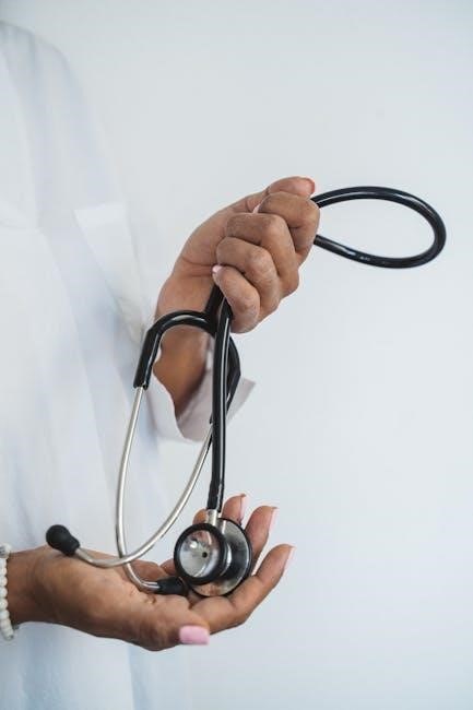
Preparation for a cranial nerve examination requires specific equipment and a structured approach. Essential tools include a torch for pupillary reactions, an ophthalmoscope for visual field assessment, and a tuning fork for hearing tests. Olfactory testing needs smell identifiers like peppermint or vanilla. A neurological hammer and gloves may also be necessary. Ensure the room is well-lit and quiet to minimize distractions. Position the patient comfortably, preferably sitting upright, to facilitate eye movement and facial assessments; Introduce yourself, explain the process, and maintain patient comfort throughout the examination. Proper preparation ensures accuracy and efficiency in evaluating cranial nerve function.
3.2 Patient Positioning and Comfort
Proper patient positioning and comfort are essential for an effective cranial nerve examination. Position the patient in a comfortable, upright sitting position with good support for their head and neck. Ensure the room is well-lit and free from distractions. For patients unable to sit, a semi-recumbent or supine position may be used. Maintain a calm and reassuring demeanor to reduce anxiety. Introduce yourself, explain the examination process, and ensure the patient is at ease. Positioning should allow easy access to the face and eyes while minimizing discomfort. This setup facilitates accurate assessment of cranial nerve function and ensures patient cooperation.
3.3 Specific Tests for Each Cranial Nerve

Each cranial nerve requires specific tests to assess its function. For Cranial Nerve I (Olfactory), test smell perception using familiar odors. For CN II (Optic), evaluate visual acuity and pupil responses. CN III (Oculomotor), CN IV (Trochlear), and CN VI (Abducens) are assessed through eye movement tests. CN V (Trigeminal) involves facial sensation and motor response. CN VII (Facial) is evaluated by testing facial muscle movements. CN VIII (Vestibulocochlear) includes hearing and balance assessments. CN IX (Glossopharyngeal) and CN X (Vagus) are tested via swallowing and gag reflex. CN XI (Accessory) assesses shoulder strength, and CN XII (Hypoglossal) evaluates tongue movement. These tests help identify deficits and correlate findings with clinical presentation.

Testing Cranial Nerves
Testing cranial nerves involves assessing sensory and motor functions. Evaluate smell, vision, eye movements, facial expressions, hearing, and swallowing. Systematic assessment ensures accurate identification of deficits and correlations with neurological conditions.
4.1 Sensory Functions (Cranial Nerves I, II, VIII)
Assessing sensory functions involves evaluating cranial nerves I (olfactory), II (optic), and VIII (vestibulocochlear). Test olfaction using scented objects, ensuring one nostril is occluded at a time. For vision, measure visual acuity and check pupillary light reflex. Evaluate hearing via tuning fork tests or audiometry. Balance assessment may include Romberg or Dix-Hallpike tests. These evaluations help identify deficits linked to neurological conditions, ensuring accurate diagnosis and early intervention. Systematic testing ensures comprehensive evaluation of sensory functions, correlating findings with specific cranial nerve pathologies.
4.2 Motor Functions (Cranial Nerves III, IV, VI, XII)
Motor functions are assessed through cranial nerves III (oculomotor), IV (trochlear), VI (abducens), and XII (hypoglossal). Evaluate extraocular movements by asking the patient to follow an object in all directions. Note any nystagmus or diplopia. For the hypoglossal nerve, observe tongue protrusion and lateral movement, checking for atrophy or fasciculations. These tests help identify motor deficits, such as ptosis, lateral gaze palsy, or tongue deviation, which may indicate specific nerve palsies or brainstem lesions; Accurate assessment ensures timely diagnosis and management of neurological conditions affecting motor cranial nerves.

Common Abnormalities and Findings
Cranial nerve examinations often reveal palsies, sensory deficits, or motor impairments. Identifying these abnormalities aids in diagnosing brainstem lesions or specific nerve damage, ensuring timely intervention.
5.1 Identifying Cranial Nerve Palsies
Cranial nerve palsies often manifest as facial droop, ptosis, or speech difficulties. These abnormalities indicate potential nerve damage or brainstem lesions. Assessing sensory and motor functions helps pinpoint affected nerves. For instance, impaired smell suggests olfactory nerve issues, while visual deficits point to optic nerve damage. Motor weaknesses, like difficulty swallowing or eye movement disorders, signal problems with specific cranial nerves. Detailed physical examination and patient history are crucial for accurate diagnosis. Testing each nerve’s function systematically ensures comprehensive evaluation and effective treatment planning.
5.2 Interpreting Sensory and Motor Deficits
Sensory deficits, such as impaired smell or vision, indicate issues with cranial nerves I, II, or VIII. Motor deficits, like facial weakness or difficulty swallowing, suggest involvement of nerves III, IV, VI, VII, or XII. Correlating these findings with patient history and imaging aids in diagnosing conditions like Lyme disease or stroke. Each deficit points to specific nerve dysfunction, guiding targeted treatments. Accurate interpretation of sensory and motor findings is essential for proper neurological assessment and effective patient care.

Interpretation of Examination Results
Accurate interpretation of cranial nerve examination results is crucial for diagnosing neurological conditions. Findings are correlated with patient history and imaging to guide targeted treatment plans effectively.
6.1 Correlating Findings with Neurological Conditions
Accurate correlation of cranial nerve examination findings with neurological conditions is essential for diagnosis. Abnormalities such as palsies or sensory deficits may indicate conditions like multiple sclerosis, Lyme disease, or stroke. For instance, optic nerve issues could suggest optic neuritis, while facial weakness may point to Bell’s palsy or brainstem lesions. Patient history and imaging are crucial for confirming diagnoses. Trigeminal nerve dysfunction might indicate trigeminal neuralgia or tumors. By linking specific deficits to known pathologies, clinicians can develop targeted treatment plans, ensuring optimal patient outcomes and addressing underlying causes effectively.
A cranial nerve examination is a vital tool in neurological assessment, aiding in the diagnosis of various conditions and guiding appropriate treatment plans for optimal patient care.
7.1 Summary of Key Points
The cranial nerve examination is essential for assessing neurological function, identifying pathologies, and guiding treatment. Each nerve has specific roles, and abnormalities can indicate conditions like Lyme disease or trauma. Proper preparation, patient positioning, and systematic testing ensure accuracy. Key findings include sensory deficits, motor weaknesses, and palsies. Correlating results with clinical history aids in diagnosis. Regular practice and updated resources enhance examination skills, ensuring effective patient care and accurate neurological assessments.